Minimally Invasive Surgery for Malignant Pectus Excavatum
Medical History
The patient is a 12-year-old girl who has had a chest wall depression since early childhood. Initially, the deformity was mild and asymptomatic, so no treatment was pursued. However, as she grew older, the depression progressively worsened, resulting in noticeable symptoms such as palpitations and shortness of breath during physical activity.
Preoperative Examination
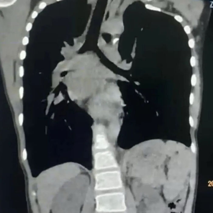
The anterior chest wall shows a severe asymmetric depression, causing marked compression of the heart, which is displaced toward the right thoracic cavity. The patient also presents with scoliosis. She is diagnosed with malignant pectus excavatum.
Surgical Overview
Three incisions, each approximately 3 cm in length, were made at the midline and on both sides of the chest wall. The chest wall first underwent pre-shaping, followed by placement of two bars to perform the Wung procedure, correcting the depression deformity. Subsequently, the Wenlin procedure was performed to further optimize the chest wall contour and achieve a more ideal appearance (a minor localized protrusion occurred during correction due to the overall rigidity and stability of the thoracic structure). The surgery proceeded smoothly and was completed within one hour. Postoperatively, the patient’s chest wall deformity was fully corrected, and normal appearance was restored.
Surgical Analysis
In clinical practice, when a depression develops in the chest wall and exerts pressure on the heart, the heart is more likely to be displaced to the left due to its normal anatomical position. Rightward displacement of the heart is extremely rare and, when present, often indicates significant cardiac compromise, putting the patient at high risk of developing serious symptoms. Based on these considerations, the chest wall deformity in this case was classified as malignant pectus excavatum, necessitating prompt surgical intervention.
The surgery for this case was highly challenging. Given the significant cardiac compression, two major intraoperative risks were present: first, the patient was at risk of sudden cardiac arrest when lying supine, necessitating immediate correction of the deformity to relieve cardiac compression and ensure patient safety. Second, due to the severe and asymmetric nature of the chest wall depression, the risk of intraoperative cardiac injury was markedly increased, and conventional surgical techniques alone would be insufficient to achieve an optimal corrective outcome.
To manage these risks, the Wung procedure was employed to correct the depression deformity. In addition, several specialized techniques, including pre-shaping, were applied. The use of these techniques significantly reduced surgical risks, allowing the procedure to be completed efficiently within a short timeframe and achieving a highly satisfactory outcome.


