
Pectus excavatum after cardiac surgery is frequently encountered in clinical practice and often raises the same question: why does it happen? This article explains the causes of pectus excavatum after cardiac surgery and why treatment can be more difficult than in ordinary pectus excavatum.
Two Types of Pectus Excavatum
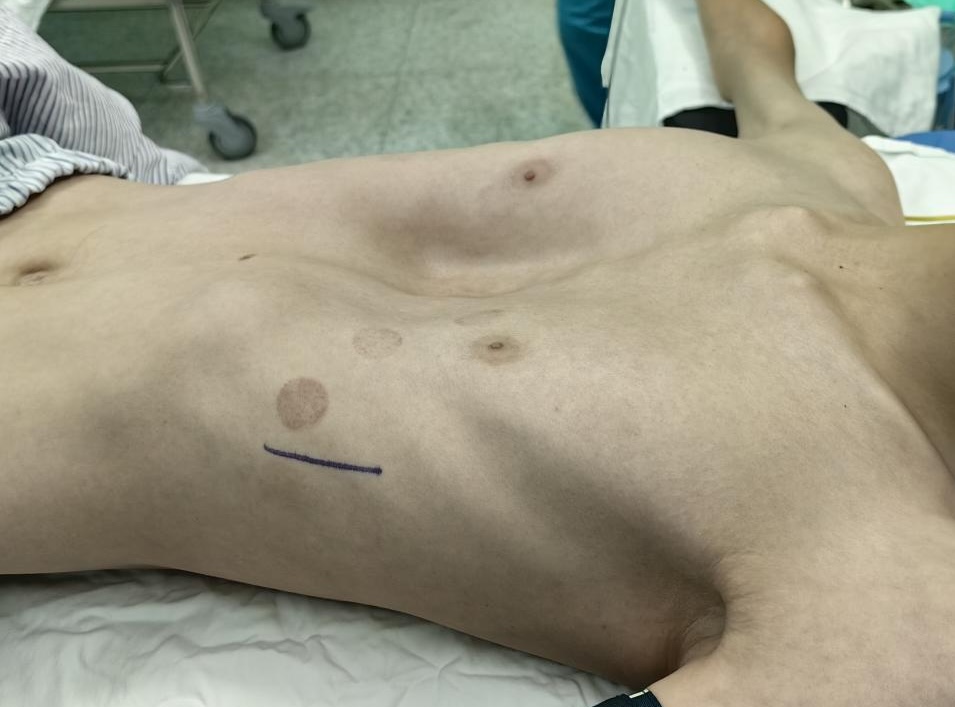

Pectus excavatum is a relatively common chest wall deformity characterized by an inward depression of the middle and lower sternum along with the adjacent costal cartilages, resulting in a funnel-shaped appearance of the anterior chest wall. The severity varies among individuals: mild cases may affect only cosmetic appearance, whereas severe cases can compress the heart and lungs and impair cardiopulmonary function.
Based on etiology, pectus excavatum can be classified into two major types: primary and secondary. The exact cause of primary pectus excavatum remains unclear. In contrast, secondary pectus excavatum may result from surgery or trauma, and pectus excavatum following cardiac surgery is the most common form of surgery-related secondary pectus excavatum.
Learn more about the risk of pectus excavatum: Is Pectus Excavatum Dangerous?
What Causes Secondary Pectus Excavatum After Cardiac Surgery?
A Complex Process Involving Multiple Factors
The development of pectus excavatum after cardiac surgery is a highly complex process that may involve multiple contributing factors. Patient-related variables such as age, skeletal development, nutritional status, and healing capacity may all play a role. However, in most cases, its occurrence is more closely associated with surgical technique and postoperative recovery.
The following three mechanisms are considered the major causes of the secondary pectus excavatum after cardiac surgery.
1. Disruption of Sternal Stability
Most cardiac procedures require a median sternotomy. During surgery, the sternum is incised and divided along the midline to expose the heart and mediastinal structures. This process inevitably compromises the native stability of the anterior chest wall, creating a predisposition for subsequent pectus excavatum.
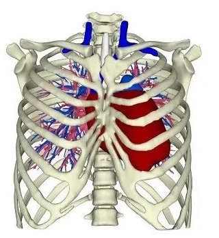
Significant gap and instability between the two sternal halves following cardiac surgery
2. Suboptimal Fixation Techniques
After completion of cardiac surgery, the sternum must be closed and stabilized. Common fixation methods include wire cerclage, tension band techniques, and rigid bar fixation. In most cases, these methods provide adequate stability. However, if the fixation technique is suboptimal or poorly matched to the patient's anatomical characteristics, the sternum may fail to maintain optimal stability during healing.
This issue is more commonly observed in infants and young children. Due to the pediatric sternum's relatively soft, highly elastic nature, achieving rigid fixation can be more challenging. As a result, postoperative sternal instability is more likely to occur in younger patients, which may explain the higher incidence of post-cardiac surgery pectus excavatum in this population.
3. Postoperative Recovery Factors
Following cardiac surgery, adhesions often develop between the sternum and mediastinal tissues. Over time, these adhesions may exert a persistent inward traction on the anterior chest wall.
In addition, postoperative complications, such as local infection or any other factors that compromise chest wall stability during recovery, may further increase the risk of developing pectus excavatum.
Is Pectus Excavatum After Cardiac Surgery More Difficult to Treat?
Yes, secondary pectus excavatum following cardiac surgery is significantly more challenging to treat than typical primary pectus excavatum.
The greatest challenge arises from dense mediastinal adhesions that develop after cardiac surgery. In minimally invasive corrective procedures such as the Nuss procedure or the Wung procedure, a metal bar must be passed through the thoracic cavity and positioned posterior to the sternum, then rotated within the chest to elevate the depressed anterior chest wall. However, the presence of adhesions can obstruct both the placement and rotation of the bar. Therefore, surgeons performing the Nuss procedure or the Wung procedure must carefully dissect and manage these adhesions, which substantially increase operative difficulty and surgical risk.
In contrast, the Wang procedure is performed outside the thoracic cavity and can therefore effectively avoid this risk, offering certain advantages in patients with secondary pectus excavatum.
Therefore, for patients with secondary pectus excavatum following cardiac surgery, the choice of surgical approach is particularly critical.
Wang Procedure: Effective Correction of Pectus Excavatum
The Wang procedure can be used to correct secondary pectus excavatum following cardiac surgery, as well as other types of pectus excavatum. This technique was introduced by Dr. Wenlin Wang in 2018 and was recognized by the National Health Commission of China in 2019 as one of the standard surgical approaches for pectus excavatum.
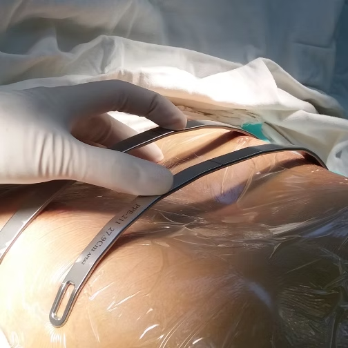
The main difference between the Wang procedure and the Nuss procedure lies in the position of the corrective bar. In the Nuss procedure, bars are inserted into the thoracic cavity, passed across the anterior surface of the heart, and then rotated within the chest to elevate the depressed anterior chest wall. In contrast, the Wang procedure places the bar on the surface of the depressed bony structures, outside the thoracic cavity. And steel wires are used to lift the sternum and costal cartilages toward the bar. Because the bar does not enter the retrosternal space, this approach avoids the intrathoracic adhesions that complicate correction after cardiac surgery and reduces the associated risk of cardiac injury. The procedure typically requires only a small incision of approximately 2 cm and does not require thoracoscopic assistance.
Learn the differences between the Wang procedure and the Nuss procedure: What are the differences between the Wang procedure and the Nuss procedure?
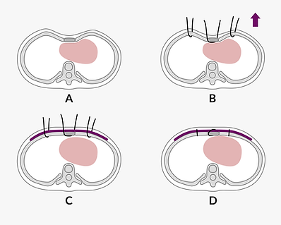
surgical technique of the Wang procedure
The Wang procedure can be applied to various types of pectus excavatum, including:
1. Primary Pectus Excavatum
-
The Wang procedure may be particularly suitable for certain types of primary pectus excavatum, including asymmetric pectus excavatum, pectus excavatum with acute angular deformity, and severe cases
- Primary pectus excavatum requiring simultaneous cardiac surgery
2.Secondary Pectus Excavatum after Cardiac Surgery
3. Persistent Pectus Excavatum after Failed Nuss Procedure or Ravitch Procedure
For more details on which cases are best suited for the Wang procedure, please refer to: What special types of pectus excavatum (PE) are suitable for the Wang procedure?
The Institute of Chest Wall Surgery (ICWS)
The Institute of Chest Wall Surgery (ICWS) is the world's largest chest wall deformity correction center. Our team is capable of treating a wide range of complex conditions, including secondary pectus excavatum following cardiac surgery. ICWS offers various treatment options, including the independently developed Wang procedure and Wung procedure, and has extensive clinical experience in chest wall reconstruction and deformity correction. To date, we have performed more than 200 surgeries for pectus excavatum after cardiac surgery, representing the largest case volume of this condition worldwide.
If you or someone you care about is affected by pectus excavatum after cardiac surgery or other chest wall deformities, you are welcome to contact ICWS to arrange a consultation.
.png)
.png)