Yes. Pectus excavatum can affect the heart, but not every patient develops cardiac dysfunction. A deeper-looking depression does not always mean a greater cardiac effect: its position and actual relationship with the heart also matter. This article explains when the heart may be displaced or compressed, which symptoms deserve attention, how doctors evaluate possible effects, and how the findings guide individualized treatment.
How Can Pectus Excavatum Affect the Heart?
Learn first: What Is Pectus Excavatum?
Pectus excavatum causes the sternum and adjacent costal cartilages to curve inward, narrowing the space between the front of the chest and the heart. The inward chest wall may displace the heart or press directly against it, but the resulting cardiac effect varies with the depression's severity, location, and anatomical relationship to the heart.
A More Extensive Depression Creates More Pressure
A deeper and broader depression leaves less space for the heart and lungs. In severe pectus excavatum, sustained pressure is more likely to limit cardiopulmonary function and produce symptoms.
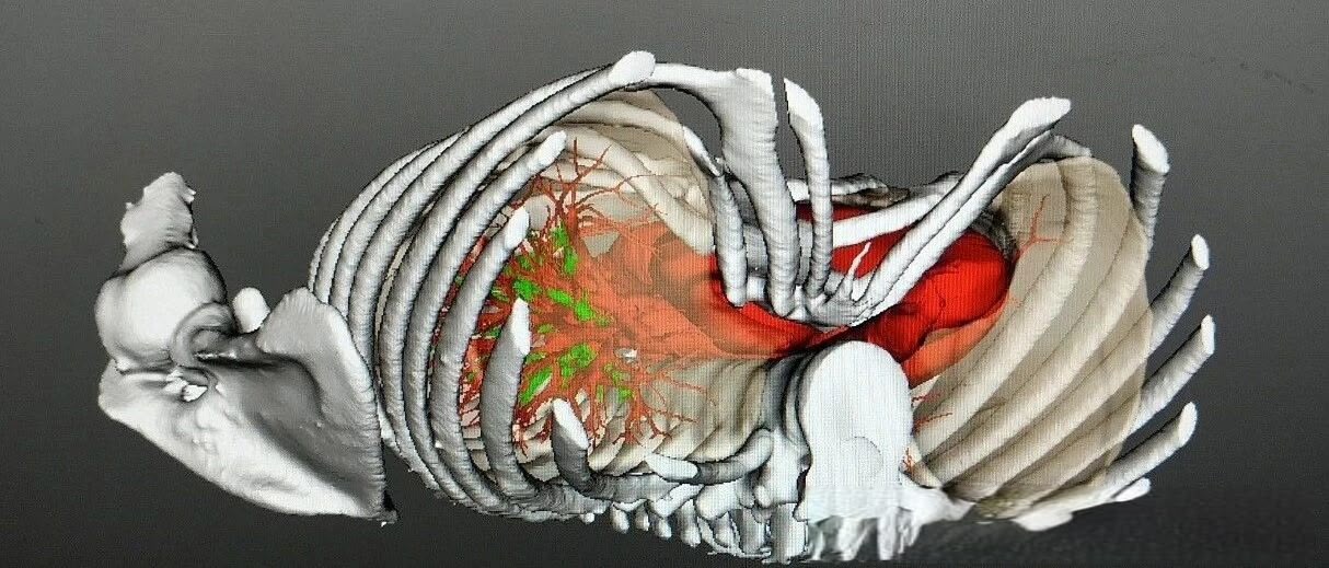
A Higher Depression Sits Closer to the Heart Base
Where the depression lies can matter as much as how deep it appears. In high-positioned pectus excavatum, the depression is close to the heart base and the points where major blood vessels enter and leave the heart. A pronounced depression in this position is more likely to affect cardiac function.
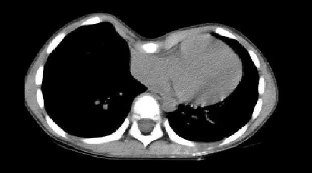
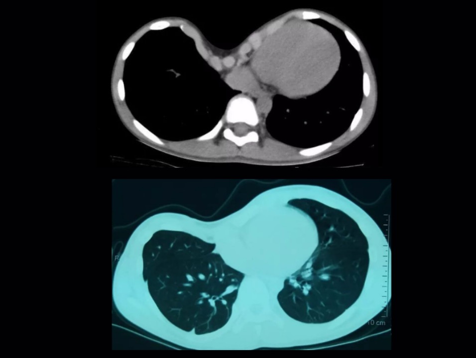
Chest CT showing cardiac compression caused by pectus excavatum
Direct Pressure Occurs When the Heart Cannot Shift
In most cases, the heart shifts toward the left side of the chest as the sternum moves inward. The resulting pressure is mainly lateral and usually has less effect on cardiac function. In malignant pectus excavatum, the heart cannot move left to avoid the depression. The anterior chest wall then presses directly against it, so cardiac dysfunction and noticeable symptoms may appear earlier.
Which Pectus Excavatum Symptoms May Suggest Cardiac Involvement?
When pectus excavatum affects cardiopulmonary function, symptoms are often more noticeable during activity and can be more prominent in severe pectus excavatum:
- Palpitations. Patients may feel their heart racing, pounding, or skipping beats.
- Chest pain or discomfort in the precordial area.
- Shortness of breath, particularly during activity. Chest tightness and breathlessness may become more noticeable after exertion.
- Reduced exercise tolerance. Usual exercise may become harder to sustain, and fatigue may appear earlier.
Mild pectus excavatum may produce no noticeable symptoms. Palpitations, chest pain, and shortness of breath may also have other cardiac or respiratory causes, while reduced exercise tolerance can reflect general fitness. A medical evaluation is therefore necessary to determine whether these symptoms are related to the chest wall deformity and whether the heart is affected.[1]
How Do Doctors Evaluate Possible Effects on the Heart?
Evaluation is tailored to the patient's symptoms, chest wall deformity pattern, and age. It may cover four complementary areas.[1-3]
Clinical Assessment
- Symptom history. The doctor records which symptoms are present, when they began, what triggers them, and how they affect activity.
- Chest wall examination. Inspection and palpation assess the depression's location, depth, extent, shape, and symmetry.
- Cardiovascular examination. The doctor listens to the heart sounds, checks whether the point of maximal impulse (PMI) is displaced, and looks for other abnormal cardiac or vascular signs.
Chest Imaging
- Chest X-ray. This provides an initial view of the chest wall's bony structure and the heart's position.
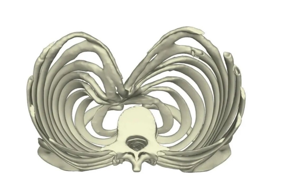
- Chest CT. Cross-sectional images show the depression and its relationship with the heart and lungs. Measurements such as the Haller Index and Wenlin Index help describe severity, while three-dimensional reconstruction may assist with complex anatomy.

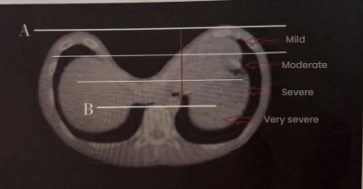
Wenlin Index for grading pectus excavatum severity
- Chest MRI. MRI may be selected for children or when further assessment of soft-tissue structures and the relationship between the sternum and heart is needed without exposure to ionizing radiation.
Cardiac Assessment
- Electrocardiography (ECG). An ECG records the heart's rhythm and electrical activity. It helps identify rhythm abnormalities that may explain palpitations or require further cardiac assessment.
- Routine transthoracic echocardiography (TTE). This cardiac ultrasound evaluates the heart's chambers, valves, size, shape, and pumping function. It helps identify structural or functional abnormalities related to compression or associated heart disease.
Respiratory Assessment
When breathlessness or reduced exercise tolerance needs further explanation, respiratory assessment may include:
- Pulmonary function testing (PFT). PFT measures lung volumes and airflow to assess whether the chest wall is restricting breathing. The results also help distinguish chest-wall restriction from other causes of breathlessness.
- Cardiopulmonary exercise testing (CPET). CPET measures exercise capacity and shows how the heart and lungs respond together during physical activity.
Together, these findings show the chest wall's internal anatomy and any measurable effect on cardiac, respiratory, or exercise function. They help the doctor determine whether symptoms relate to the deformity and whether treatment is needed.
How Does ICWS Plan Pectus Excavatum Treatment?
At the Institute of Chest Wall Surgery (ICWS), treatment is tailored to the patient’s chest wall anatomy and needs. Our team considers symptoms, the pattern and severity of the depression, documented effects on the heart or lungs, age, and the patient's physical and psychological concerns.
ICWS offers two minimally invasive pectus excavatum surgery approaches. Both reduce surgical trauma and shorten postoperative recovery, while their operative principles and most appropriate patient groups differ:
- Wang procedure. It can be applied to young children, including patients under 5. The corrective bar is placed on the outer surface of the depressed bony chest wall, without passing through the chest cavity, thereby avoiding potential cardiac injury. Central fixation does not restrict normal bone development.

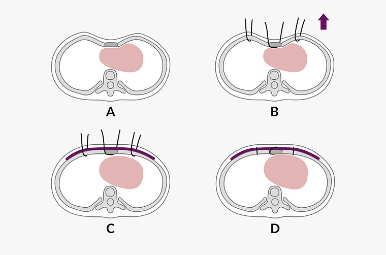
Surgical technique illustration of the Wang procedure
- Wung procedure. Pre-shaped bars are placed inside the chest cavity to lift the depression. A guiding catheter supports safer bar placement, while refined fixation improves bar stability. Pre-shaping helps address the greater chest-wall rigidity often seen in adults, making the deformity easier to correct.
LEARN MORE: What is Pre-shaping?
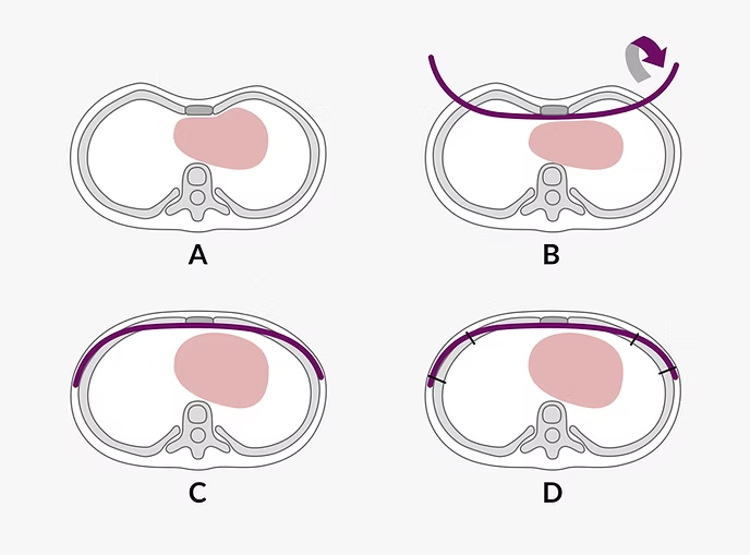
Surgical technique illustration of the Wung procedure
ICWS specializes in chest wall disorders and has spent more than 15 years researching and treating these conditions. We have performed over 10,000 chest wall surgeries, including complex deformities and revision cases. If you or your child has pectus excavatum and are concerned about possible heart problems, request an appointment for an individual assessment and treatment discussion.
FAQ
Can Pectus Excavatum Cause Chest Pain?
Yes. Chest pain or discomfort in the precordial area is one of the possible pectus excavatum symptoms. Because chest pain may also have cardiac, respiratory, or musculoskeletal causes, a medical evaluation is needed to identify the source.
Can Pectus Excavatum Cause Heart Problems Without Obvious Symptoms?
Yes. The heart may maintain its function despite external compression, so cardiac compression can be present before noticeable symptoms or detectable functional abnormalities develop. The absence of symptoms does not rule out cardiac involvement.
Why Can Pectus Excavatum Appear After Cardiac Surgery?
Pectus excavatum after cardiac surgery is a form of secondary pectus excavatum. Many cardiac procedures require a median sternotomy, which divides the sternum and reduces the anterior chest wall's natural stability. If sternal fixation does not maintain sufficient support during healing, an inward depression may develop. Postoperative adhesions may also pull the chest wall inward, while infection or other recovery-related problems can further compromise stability. Dense adhesions can make later corrective surgery more difficult.
References
[1] Dunning J, Burdett C, Child A, et al. The pectus care guidelines: best practice consensus guidelines from the joint specialist societies SCTS/MF/CWIG/BOA/BAPS for the treatment of patients with pectus abnormalities. European Journal of Cardio-Thoracic Surgery. 2024;66(1):ezae166.
[2] Coorens NA, Janssen N, Daemen JHT, et al. Advancements in preoperative imaging of pectus excavatum: a comprehensive review. Journal of Thoracic Disease. 2024;16(1):696-707.
[3] Janssen N, Daemen JHT, van Polen EJ, et al. Pectus Excavatum: Consensus and Controversies in Clinical Practice. The Annals of Thoracic Surgery. 2023;116(1):191-199.
`